
What Does “Return to Sport” Actually Mean?
Tue, 29 Sep 2026

Follow the stories of academics and their research expeditions
Principles & Biophysical Mechanisms: How It Works
Therapeutic ultrasound operates by converting electrical energy into high-frequency acoustic energy via the reverse piezoelectric effect. This process occurs inside an applicator head containing a synthetic ceramic crystal (such as lead zirconate titanate) that expands and contracts in response to an alternating electrical current.
These rapid crystal oscillations generate longitudinal sound waves that travel into biological tissue, alternating between phases of high pressure (compression) and low pressure (rarefaction) (Miller et al., 2012).
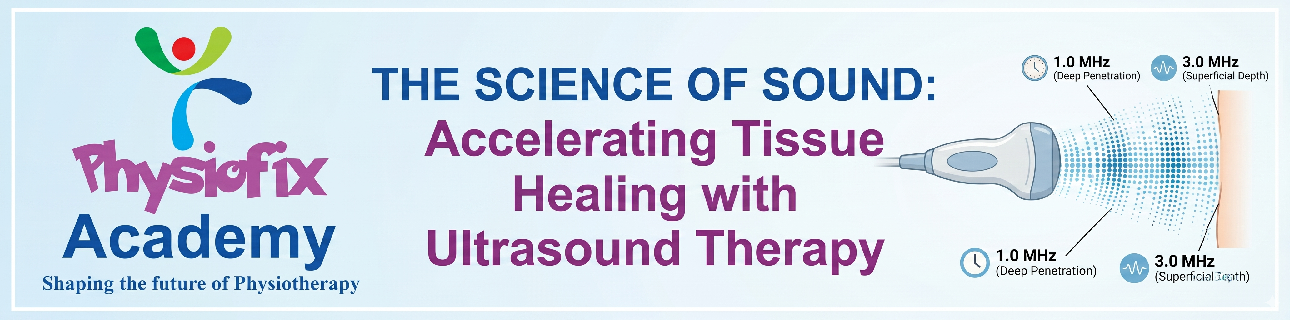
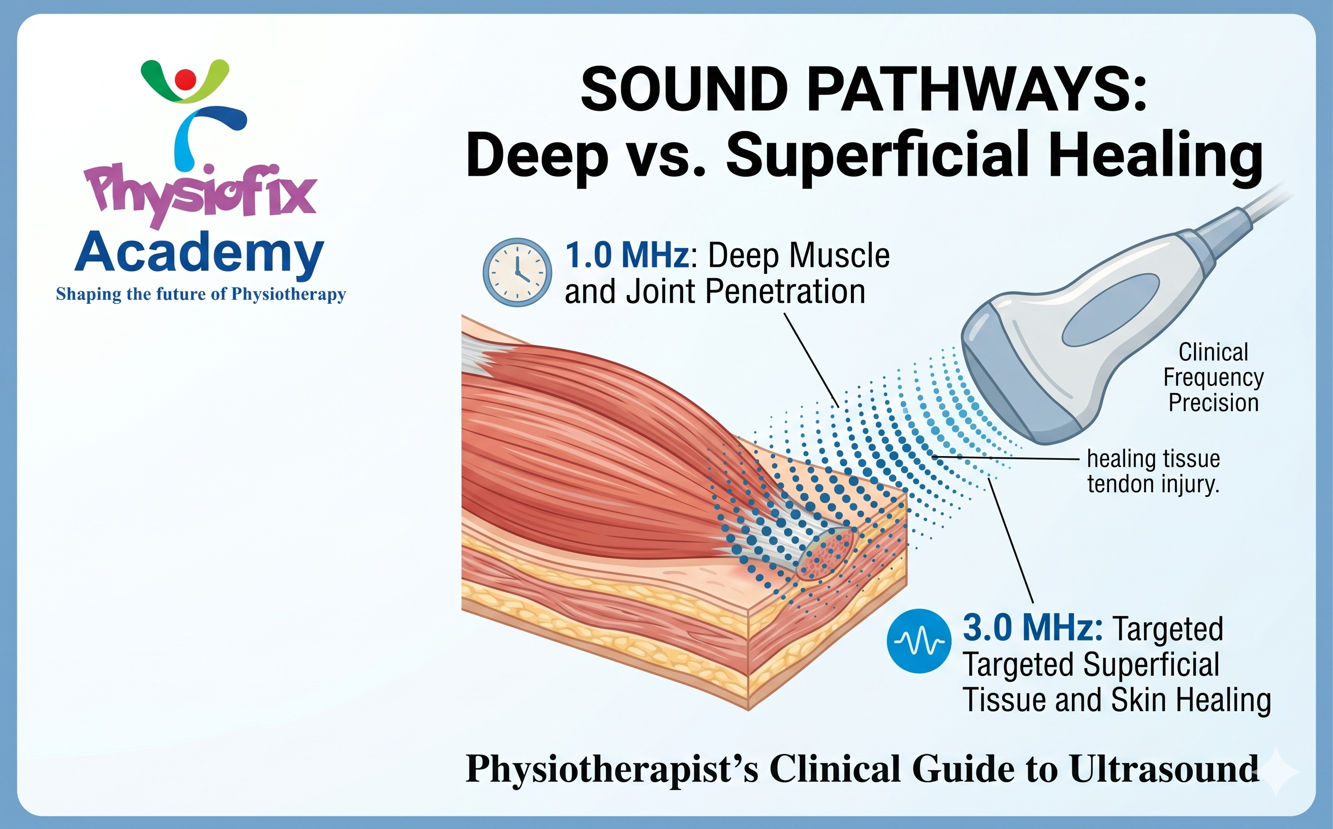
Frequency: Determines the depth of penetration. A frequency of 1.0 MHz penetrates deeply (up to 5 cm to target deep muscles and joint capsules), while 3.0 MHz is absorbed superficially (up to 1.5 cm to target skin, superficial tendons, and fascia) (Bader et al., 2024).
Waveform Mode: Dictates whether energy delivery is uninterrupted (Continuous Mode) to generate thermal accumulation, or interrupted (Pulsed Mode) to favor purely mechanical, non-thermal effects.
The transition of ultrasound from industrial use to clinical medicine followed a clear timeline:
1930s–1940s (Early Exploration): Medical researchers began experimenting with ultrasound's biological effects, primarily focusing on intense tissue heating and localized, highly destructive interventions in the brain (such as treating Parkinson's disease by creating targeted lesions) (ter Haar & Coussios, 2007; Miller et al., 2012).
1950s (The Pivot to Low-Power): Rather than destroying tissue, researchers developed low-power applications (around $1.0\text{ MHz}$) specifically designed to manage inflammatory musculoskeletal conditions like tendinitis or bursitis safely (Miller et al., 2012).
1970s (Widespread Physical Therapy Adoption): Ultrasound became a standard electrophysical agent in physical therapy clinics worldwide for pain relief and soft tissue restoration (Robertson & Baker, 2001; Miller et al., 2012).
Modern Era (Expanded Scope): Beyond standard therapeutic heating and cellular repair, contemporary physical therapists also utilize Point-of-Care Ultrasound (POCUS) for rehabilitative muscle morphology imaging, diagnostic tendinopathy screens, and guiding dry-needling procedures (Whittaker et al., 2019).
The biological responses triggered by therapeutic ultrasound are divided into thermal and non-thermal categories:
When ultrasound waves pass through tissue, friction between vibrating molecules converts acoustic energy into heat (Miller et al., 2012). Tissues rich in collagen—such as tendons, ligaments, joint capsules, and fascia—have high absorption coefficients and heat rapidly (de Jesus et al., 2019). Clinical outcomes include:
Accelerated local metabolic rate and enhanced blood flow.
Increased collagen extensibility, which improves range of motion when combined with stretching.
Altered nerve conduction velocities, providing temporary pain relief and reducing muscle spasms (Bader et al., 2024).
Non-thermal responses occur regardless of thermal accumulation and are driven by two main physical processes:
Cavitation: The formation, growth, and pulsation of microscopic gas bubbles within extracellular fluid due to pressure changes (Miller et al., 2012). Stable cavitation alters cell membrane permeability without destroying tissue.
Acoustic Streaming / Micro-streaming: The localized, one-directional movement of fluids around these pulsating bubbles and cell membranes (Bader et al., 2024).
This mechanical stress deforms cell membranes, opening calcium channels and stimulating cellular activity. This activity accelerates the degranulation of mast cells, enhances macrophage performance, and boosts fibroblast synthesis of collagen to accelerate tissue repair (Miller et al., 2012).
To achieve reproducible therapeutic effects, clinicians must transition away from arbitrary "10-minute treatments" and calculate specific energy profiles. Therapeutic ultrasound dosing relies heavily on three key variables: the Effective Radiation Area (ERA), the Beam Non-uniformity Ratio (BNR), and Total Cumulative Energy (de Jesus et al., 2019).
Historically, ultrasound has faced criticism regarding its standalone efficacy because it is frequently used as a passive, unmeasured "add-on" at the end of a session (Robertson & Baker, 2001). To use ultrasound as an active, effective clinical tool, practitioners must treat it as a targeted primer for mechanotransduction rather than a passive modality.
Thermal ultrasound increases tissue elasticity for only 5–10 minutes post-application before tissue temperature cools back to baseline.
The Adjunct Pitfall: Applying ultrasound, wiping off the gel, and sending the patient home.
The Monotherapy Strategy: Administer continuous thermal ultrasound immediately before or simultaneously with manual joint mobilizations, dynamic stretching, and eccentric loading exercises to structurally re-align newly pliable collagen fibers.
Therapists must align the parameter selection with the specific phase of tissue healing rather than applying a generic setting:
[Acute Phase]      --> Pulsed Mode (20%)     --> Non-Thermal / Cellular Permeability
[Subacute Phase]   --> Pulsed Mode (50%)     --> Transition Phase / Controlled Mechanics
[Chronic Phase]    --> Continuous Mode (100%)--> Deep Tissue Heating / Collagen Extensibility
Because air reflects nearly 100% of acoustic energy, maintaining an optimal acoustic interface is critical. Therapists must use sufficient conductive gel, keep the transducer perfectly perpendicular to the skin, and move the soundhead at a slow, deliberate speed (approx 4/s) strictly within an area no larger than 2 to 3 times the ERA (de Jesus et al., 2019). This approach avoids under-dosing and ensures that the target tissue receives the exact calculated energy density required to stimulate structural adaptation.
Bader, K. B., Padilla, F., Haworth, K. J., Ellens, N., Dalecki, D., Miller, Douglas L., & Wear, K. A. (2024). Overview of Therapeutic Ultrasound Applications and Safety Considerations: 2024 Update. Journal of Ultrasound in Medicine, 44(2), 381–433.
Cited by: 47
de Jesus, J. F., de Albuquerque, T. A. B., Shimba, L. Girardi, Bryk, F. F., Cook, J., & Pinfildi, C. E. (2019). High-energy dose of therapeutic ultrasound in the treatment of patellar tendinopathy: protocol of a randomized placebo-controlled clinical trial. BMC Musculoskeletal Disorders, 20(1).
Miller, Douglas L., Smith, N. B., Bailey, M. R., Czarnota, G. J., Hynynen, K., & Makin, I. R. S. (2012). Overview of Therapeutic Ultrasound Applications and Safety Considerations. Journal of Ultrasound in Medicine, 31(4), 623–634.
Cited by: 1001
Robertson, V. J., & Baker, K. G. (2001). A Review of Therapeutic Ultrasound: Effectiveness Studies. Physical Therapy, 81(7), 1339–1350.
Cited by: 745
ter Haar, G., & Coussios, C. (2007). High intensity focused ultrasound: Physical principles and devices. International Journal of Hyperthermia, 23(2), 89–104.
Cited by: 1076
Whittaker, J. L., Ellis, R., Hodges, P. W., O'Sullivan, C., Hides, J., Fernandez-Carnero, S., Arias-Buria, J. L., Teyhen, D. S., & Stokes, M. J. (2019). Imaging with ultrasound in physical therapy: What is the PT’s scope of practice? A competency-based educational model and training recommendations. British Journal of Sports Medicine, 53(23), 1447–1453.
Cited by: 160
Tue, 29 Sep 2026

Master physiotherapy from the comfort of your home with expert-led courses

Leave a comment